Models
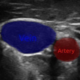
Jugular carotid ultrasound segmentation model
Jugular carotid ultrasound segmentation model
ID: jugular-carotid-ultrasound-segmentation-model
Downloads: 11
Authors: Erik Smistad
Copyrights: SINTEF
Licenses:
CC BY 4.0
Breast Tumour Segmentation Model (H2G-Net)
A neural network model for segmenting breast cancer tumours from whole-slide images using H2G-Net: https://github.com/andreped/H2G-Net
ID: breast-tumour-segmentation-model
Downloads: 198
Authors: André Pedersen
Copyrights: NTNU
Licenses:
CC BY 4.0

BACH Classification Model
Patch-wise image classification model trained on data from the 2018 breast cancer histology (BACH) challenge.
ID: bach-model
Downloads: 144
Authors: André Pedersen
Copyrights: NTNU
Licenses:
CC BY 4.0
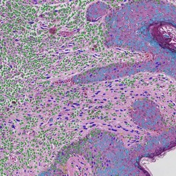
Nuclei segmentation model
Patch-wise segmentation of cell nuclei at 20X magnification using a lightweight U-Net. Trained on PanNuke dataset.
ID: nuclei-segmentation-model
Downloads: 301
Authors: André Pedersen
Copyrights: NTNU
Licenses:
CC BY 4.0
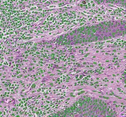
Nuclei Classfication Model (PanNuke, HoVerNet)
This model segments cell nuclei into one of 5 different classes: Neoplastic, Inflammatory, Connective, Dead, Non-Neoplastic Epithelial. It is trained on the PanNuke dataset using the HoVerNet architecture. The model is part of the publication "A Pragmatic Machine Learning Approach to Quantify Tumor-Infiltrating Lymphocytes in Whole Slide Images" Cancers 2022 https://doi.org/10.3390/cancers14122974
ID: nuclei-classification-model-hovernet-pannuke
Downloads: 84
Authors: Nikita Shvetsov, Morten Grønnesby, Edvard Pedersen, Kajsa Møllersen, Lill-Tove Rasmussen Busund, Ruth Schwienbacher, Lars Ailo Bongo, Thomas Karsten Kilvaer
Copyrights: UiT - The Arctic University of Norway
Licenses:
CC SA-BY-NC 4.0

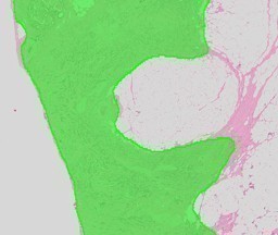
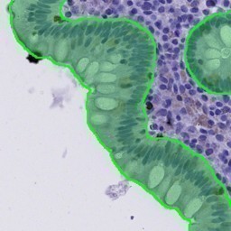
Colon epithelium segmentation model (CD3)
Model for segmentation of epithelial cells in CD3-stained biopsies of colonic mucosa of active and inactivate inflammatory bowel disease. For more info see the article "Code-Free Development and Deployment of Deep Segmentation Models for Digital Pathology" Frontiers in Medicine 2022 https://www.frontiersin.org/articles/10.3389/fmed.2021.816281/full and also the webpage https://github.com/andreped/NoCodeSeg.
ID: colon-epithelium-segmentation-cd3-model
Downloads: 59
Authors: Henrik Sahlin Pettersen, Ilya Belevich, Elin Synnøve Røyset, Erik Smistad, Melanie Rae Simpson, Eija Jokitalo, Ingerid Reinertsen, Ingunn Bakke, André Pedersen
Copyrights: NTNU, St. Olavs Hospital, SINTEF, University of Helsinki
Licenses:
GPL 2.0
Colon epithelium segmentation model (HE)
Model for segmentation of epithelial cells in HE-stained biopsies of colonic mucosa of active and inactivate inflammatory bowel disease. For more info see the article "Code-Free Development and Deployment of Deep Segmentation Models for Digital Pathology" Frontiers in Medicine 2022 https://www.frontiersin.org/articles/10.3389/fmed.2021.816281/full and also the webpage https://github.com/andreped/NoCodeSeg.
ID: colon-epithelium-segmentation-he-model
Downloads: 82
Authors: Henrik Sahlin Pettersen, Ilya Belevich, Elin Synnøve Røyset, Erik Smistad, Melanie Rae Simpson, Eija Jokitalo, Ingerid Reinertsen, Ingunn Bakke, André Pedersen
Copyrights: NTNU, St. Olavs Hospital, SINTEF, University of Helsinki
Licenses:
GPL 2.0
Brain MRI Segmentation Model
A 3D neural network which segments the brain from an MRI image.
ID: brain-mri-segmentation-model
Downloads: 11
Authors: David Bouget
Copyrights: SINTEF
Licenses:
BSD 2-Clause
Brain Tumor Segmentation Model
Segmentation model for high-grade glioblastoma brain tumors from T1 MRI volumes using AGU-Net. The method is described in the article "Preoperative Brain Tumor Imaging: Models and Software for Segmentation and Standardized Reporting" Frontiers in Neurology 2022: https://doi.org/10.3389/fneur.2022.932219
ID: brain-mri-hgg-tumor-segmentation-model
Downloads: 4
Authors: David Bouget, André Pedersen, Asgeir S. Jakola, Vasileios Kavouridis, Kyrre E. Emblem, Roelant S. Eijgelaar, Ivar Kommers, Hilko Ardon, Frederik Barkhof, Lorenzo Bello, Mitchel S. Berger, Marco Conti Nibali, Julia Furtner, Shawn Hervey-Jumper, Albert J. S. Idema, Barbara Kiesel, Alfred Kloet, Emmanuel Mandonnet, Domenique M. J. Müller, Pierre A. Robe, Marco Rossi, Tommaso Sciortino, Wimar A. Van den Brink, Michiel Wagemakers, Georg Widhalm, Marnix G. Witte, Aeilko H. Zwinderman, Philip C. De Witt Hamer, Ole Solheim, Ingerid Reinertsen
Copyrights: SINTEF, NTNU, St. Olavs Hospital
Licenses:
BSD 2-Clause
Cardiac LV Ultrasound Segmentation Light Model
A lightweight UNet model (~2 million parameters) which segments the left ventricle (LV), myocardium and left atrium (LA) in cardiac ultrasound images. Trained on the 500 patient CAMUS dataset.
ID: cardiac-lv-ultrasound-segmentation-light-model
Downloads: 10
Authors: Gilles Van De Vyver, Erik Smistad
Copyrights: NTNU
Licenses:
CC BY 4.0
Cardiac LV Ultrasound Segmentation nnU-net Model
A heavy nnU-Net model (~33 million parameters) which segments the left ventricle (LV), myocardium and left atrium (LA) in cardiac ultrasound images. Trained on the 500 patient CAMUS dataset.
ID: cardiac-lv-ultrasound-segmentation-nnunet-model
Downloads: 3
Authors: Gilles Van De Vyver, Erik Smistad
Copyrights: NTNU
Licenses:
CC BY 4.0
Cardiac LV Ultrasound Segmentation Graph Model
A graph convolutional network model which segments the left ventricle (LV), myocardium and left atrium (LA) in cardiac ultrasound images. This model was trained on the 500 patient CAMUS dataset. The network is descibed in the article "Towards Robust Cardiac Segmentation using Graph Convolutional Networks" Van De Vyver et al. 2023 https://arxiv.org/pdf/2310.01210
ID: cardiac-lv-ultrasound-segmentation-graph-model
Downloads: 2
Authors: Gilles Van De Vyver, Erik Smistad
Copyrights: NTNU
Licenses:
CC BY 4.0